Request Demo
Last update 23 Jan 2025
Biotech Research & Innovation Centre
Last update 23 Jan 2025
Overview
Tags
Infectious Diseases
Small molecule drug
Disease domain score
A glimpse into the focused therapeutic areas
No Data
Technology Platform
Most used technologies in drug development
No Data
Targets
Most frequently developed targets
No Data
| Disease Domain | Count |
|---|---|
| Infectious Diseases | 1 |
| Top 5 Drug Type | Count |
|---|---|
| Small molecule drug | 1 |
Related
1
Drugs associated with Biotech Research & Innovation CentreTarget- |
Mechanism- |
Active Org. |
Originator Org. |
Active Indication |
Inactive Indication- |
Drug Highest PhasePreclinical |
First Approval Ctry. / Loc.- |
First Approval Date- |
100 Clinical Results associated with Biotech Research & Innovation Centre
Login to view more data
0 Patents (Medical) associated with Biotech Research & Innovation Centre
Login to view more data
85
Literatures (Medical) associated with Biotech Research & Innovation Centre01 Dec 2024·Acta Biomaterialia
Machine Learning identifies remodeling patterns in human lung extracellular matrix
Article
Author: Emerson, Monica J ; Reuten, Raphael ; Dahl, Anders B ; Lund, Thomas K ; Erler, Janine T ; Brøchner, Christian B ; Madsen, Chris D ; Jensen, Thomas H L ; Willacy, Oliver ; Mayorca-Guiliani, Alejandro E
20 Jul 2023·Molecular cell
Chromatin regulation of transcriptional enhancers and cell fate by the Sotos syndrome gene NSD1.
Article
Author: Lin, Yuan ; Liu, Dingyu ; Sun, Zhen ; Hedehus, Lin ; Vierbuchen, Thomas ; Sawyers, Charles L ; Huang, Chang ; Islam, Mohammed T ; Helin, Kristian ; Koche, Richard
02 Jun 2023·Cancer Research
Abstract A047: Panorama of complex structural variants in primary localized prostate cancer
Author: Girma, Etsehiwot ; Papenfuss, Tony ; Li, Yilong ; Helin, Kristian ; Reimand, Jüri ; Feran, Breon ; Favero, Francesco ; Weischenfeldt, Joachim ; Olsen, André
3
News (Medical) associated with Biotech Research & Innovation Centre05 Dec 2023
The Biotech Innovation Organization on Tuesday named Amicus Therapeutics co-founder and longtime leader John Crowley as its new CEO, taking over a spot interim chief Rachel King has held for the past 14 months.BIOs decision to name Crowley, who will assume the role effective March 4, as permanent CEO comes just over a year after the tumultuous exit of Michelle McMurry-Heath, who left the organization after reportedly clashing with its board amid lagging staff morale and financial problems.Crowley will take the reins at a time when the industry group is challenging the U.S. government on at least two fronts: The newly granted drug pricing power bestowed to Medicareunder the Inflation Reduction Act, and the Federal Trade Commissions more aggressive stance toward biopharmaceutical deals.Crowley has experience not only lobbying for the industry before Congress, but advocating for rare disease research and leading one of its companies, Amicus, for nearly two decades.In a statement, Ted Love, BIOs board chair and the former CEO of Global Blood Therapeutics, praised Crowleys unwavering commitment to patients.He possesses a lifelong dedication to service, whether fighting for his country, wisely negotiating on behalf of our companies in Congress, or advocating in hospitals on behalf of his children and patients with rare disease, Love said in a statement.Crowley pivoted from financial consulting to the pharmaceutical industry after two of his children were born with a rare disorder called Pompe disease. After working in a management role at Bristol Myers Squibb, he ran a biotech known as Novazyme, which successfully developed a treatment for Pompe now marketed by Sanofi. The story was recounted in a book and later, the motion picture Extraordinary Measures.He joined Amicus in 2005 and served as its CEO through 2022 when he transitioned to a role as the companys executive chairman. During that time the New Jersey-based company won approval for two rare-disease drugs, Galafold for Fabry disease and Pombiliti for Pompe.That journey wasnt without its setbacks. The Food and Drug Administration hesitated to approve Galafoldapproval because of limited data, but later reversed course and cleared it, a turnaround linked to a meeting between Crowley and former President Donald Trump.Amicus also still isnt turning a profit. In 2022, it recorded a lossof $237 million on $329 million in product revenue. '
Executive ChangeDrug ApprovalFinancial Statement
05 Jul 2023
More than 10 urgent visits to the bathroom a day due to diarrhea can make it virtually impossible to lead a normal life. But new research can help doctors diagnose bile acid diarrhea and find the right treatment.
Most people have at some point in their life suffered an intestinal infection or food poisoning forcing them to stay close to the bathroom. It is very uncomfortable. Most of the time, though, it passes quickly.
But around 60,000-100,000 Danes suffer from a form of chronic diarrhea called bile acid malabsorption or bile acid diarrhea.
It is a chronic condition characterised by frequent and sudden diarrhea more than 10 times a day. Even though the disease is not life-threatening, it can seriously affect the patient's everyday life, especially their social life, and be extremely disabling.
"You have to rush to the bathroom several times a day. Therefore, keeping a job or maintaining social relations can be difficult, and a lot of people isolate themselves. The disease controls their life," says Professor Jesper Bøje Andersen from the Biotech Research & Innovation Centre.
He and his research group and clinical cooperation partners at Herlev and Gentofte Hospital headed by Professor and Consultant Doctor Filip Krag Knop are responsible for a new study, which provides new ways of diagnosing bile acid diarrhea and identifying the most effective treatment for the individual patient.
"A lot of people with chronic diarrhea don't realise that they suffer from bile acid diarrhea and what has caused it. This is a result of lack of knowledge among healthcare workers and the relatively complex and expensive -- and for the patient difficult -- process of diagnosing the disease," says Filip Krag Knop.
Jesper Bøje Andersen adds:
"We have developed a new concept which may be used to diagnose the disease based on a simple blood sample. Today, diagnostics involves radiopharmaceuticals, which means that there is a radiation risk. The process is not necessarily dangerous, but unpleasant and arduous, and not all countries in the world support the method, including the US."
The new method means that doctors should be able to determine whether the patient has bile acid diarrhea based on a simple blood sample. They focus on molecules known as metabolites in the blood.
"A blood sample contains lots of different metabolites. Right now we are able to identify almost 1,300 different metabolites, and around a handful of these can be used to diagnose bile acid diarrhea. The metabolites of bile acid diarrhea patients form a particular pattern that makes them recognisable," says Jesper Bøje Andersen.
Which treatment?
The researchers analysed blood samples from 50 patients and they quickly realised that the samples -- and patients -- could be divided into two groups.
"First, we did not understand why. All the blood samples had been taken before treatment, typically at the time of diagnosis," says Jesper Bøje Andersen.
The patients then participated in a randomised clinical study at the Center for Clinical Metabolic Research at Herlev and Gentofte Hospital. Here the doctors studied the effect of two different treatments: the conventional treatment involving bile acid sequestrant colesevelam and a new treatment involving liraglutide, which is normally used to treat type 2 diabetes and severe overweight.
"What is interesting is that the metabolites in the patients' blood divided them into two groups: one that responds well to colesevelam and one that responds well to liraglutide. This suggests that we should be able to say which treatment is the most effective by analysing the patient's blood at the time of diagnosis," says Jesper Bøje Andersen.
The clinical study showed that colesevelam treatment eased the bile acid diarrhea symptoms of 50 per cent of the patients, while liraglutide treatment eased the symptoms of 77 per cent of the patients.
Jesper Bøje Andersen, Filip Krag Knop and their research groups hope the new study will benefit the 60,000-100,000 Danes who suffer from bile acid diarrhea.
The majority of cases of bile acid diarrhea is diagnosed at a very late stage or never diagnosed at all.
"Around 40 per cent of the patients suffer from this condition for up to five years before it is diagnosed. Of course, this may be because they do not realise that it is a disease and that it can be treated. But it may also be because chronic diarrhea is a tabooed disease," says Filip Krag Knop.
27 Jul 2022
How do you detect a dangerous cancer if you do not know exactly what to look for or where? New research into biliary tract cancer can pave the way for early detection of the deadliest cancers.
One of the deadliest forms of cancer is biliary tract cancer. Only one in three patients diagnosed with the disease is operable. The rest must settle for life-sustaining treatment.
The reason why this cancer is so deadly is that it is difficult to diagnose, and therefore, most patients are not diagnosed with the disease until after the cancer has had time to spread.
Nevertheless, new research from the University of Copenhagen can pave the way for early detection of biliary tract cancer and other serious cancers.
"Our study shows that biliary tract cancer causes the immune cells to change behaviour, resulting in a unique expression of microRNA molecules in the patient's blood. These changes enable us to diagnose biliary tract cancer much earlier than with existing tests," says Associate Professor Jesper Bøje Andersen. He is head of the group of researchers from the Biotech Research & Innovation Centre at the University of Copenhagen who are responsible for the new study.
"Sometimes tumours, including the ones you find in the biliary tract, differ considerably, and developing a comprehensive measure for these tumours can therefore be difficult. But one thing all cancers have in common is the fact that they affect the immune system," says PhD Dan Høgdall, who is first author of the study and a doctor at the Department of Oncology at Herlev and Gentofte Hospital. He adds:
"We need to focus attention on how cancer affects the body as a whole instead of focussing solely on the cancer cells. Among other things, such a broad approach has paved the way for brand new treatments involving immunotherapy, which is targeted at the immune cells instead of the cancer cells. Adopting a broad approach can also provide us with important knowledge about early diagnostics."
Cancer causes the immune cells to change behaviour
The researchers have examined more than 200 blood samples from people with and without biliary tract cancer. They have analysed the cells in the blood, a large part of which were immune cells. More specifically, they have conducted microRNA analyses. MicroRNA is a group of genes, which play a key part in the complex development of the human genome.
"By comparing the different levels of microRNA in the blood, we identified four microRNAs, and this enabled us to distinguish patients with biliary tract cancer from healthy participants. Other types of blood analyses were unable to do that. All in all, the data indicates that microRNAs change in patients with biliary tract cancer," says Jesper Bøje Andersen.
The new study is not the first to research cancer and the immune system, but it is the first to do so in relation to biliary tract cancer.
"The research method is also new. We look at the blood as a whole and thus at all the cells, which largely consist of immune cells. A lot of research seeks to identify methods for early detection of cancer. But it is like looking for a needle in a haystack, as the goal is to find the tumours while they are still very small. The idea behind this approach is to look not for the needle, but for small changes in the haystack," explains Dan Høgdall.
Even though the researchers have completed the study, it will be a while before the new method can be used to diagnose patients.
"This is basic research, which means that it will take some time. But it does suggest that it makes sense to look at the systemic impact of cancer. It will require more in-depth research, though," he says.
Biliary tract cancer
Immunotherapy
100 Deals associated with Biotech Research & Innovation Centre
Login to view more data
100 Translational Medicine associated with Biotech Research & Innovation Centre
Login to view more data
Corporation Tree
Boost your research with our corporation tree data.
login
or

Pipeline
Pipeline Snapshot as of 24 Feb 2025
The statistics for drugs in the Pipeline is the current organization and its subsidiaries are counted as organizations,Early Phase 1 is incorporated into Phase 1, Phase 1/2 is incorporated into phase 2, and phase 2/3 is incorporated into phase 3
Preclinical
1
Login to view more data
Current Projects
| Drug(Targets) | Indications | Global Highest Phase |
|---|---|---|
LB 10827 | invasive Streptococcus pneumoniae infection More | Preclinical |
Login to view more data
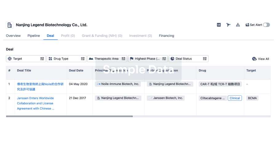
Deal
Boost your decision using our deal data.
login
or
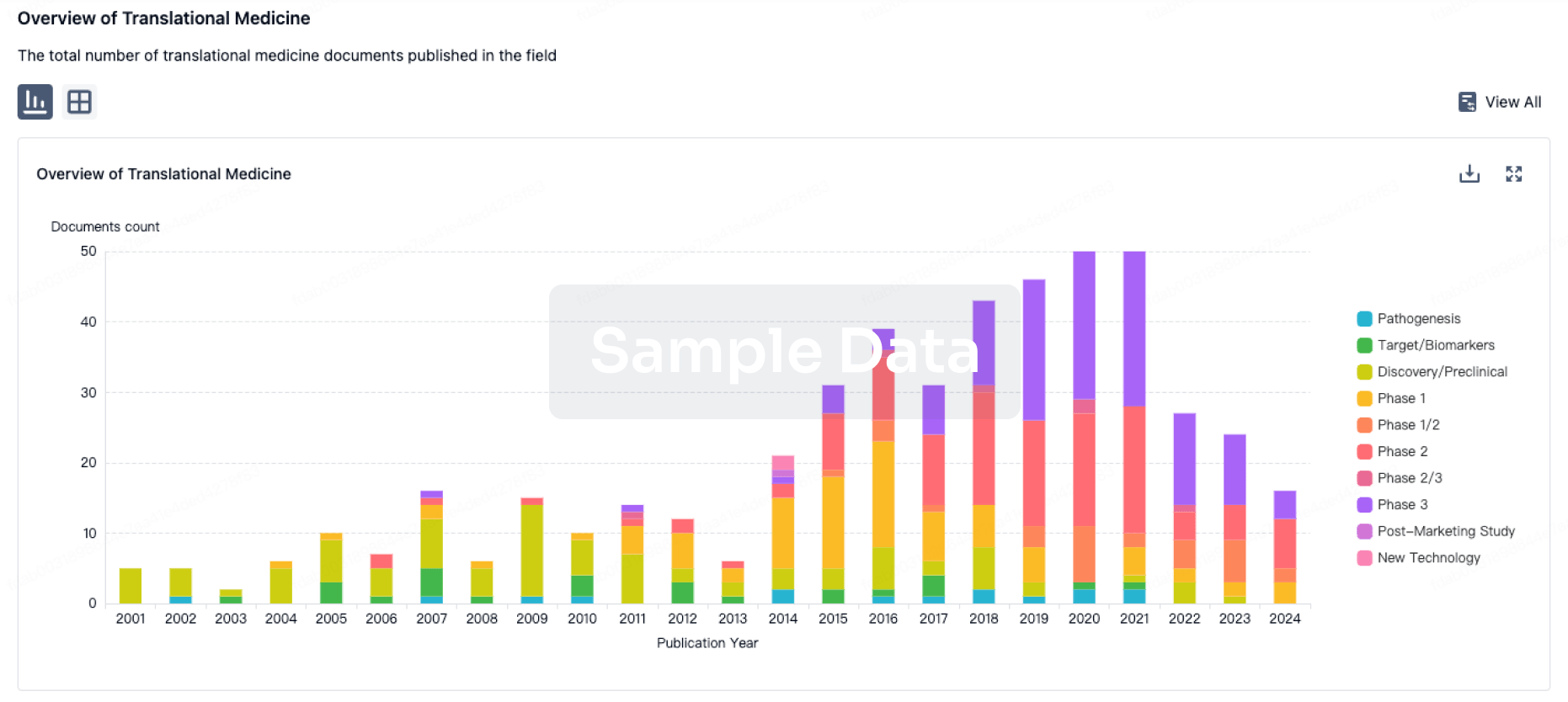
Translational Medicine
Boost your research with our translational medicine data.
login
or
Profit
Explore the financial positions of over 360K organizations with Synapse.
login
or

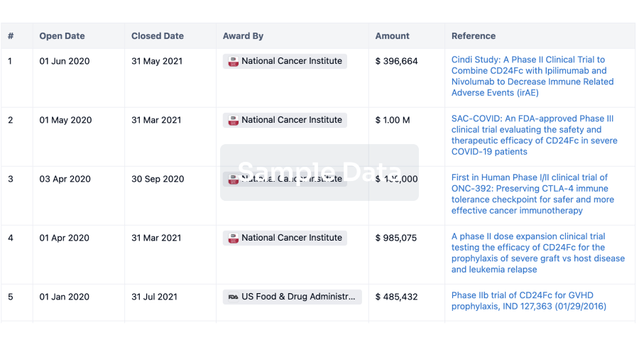
Grant & Funding(NIH)
Access more than 2 million grant and funding information to elevate your research journey.
login
or
Investment
Gain insights on the latest company investments from start-ups to established corporations.
login
or
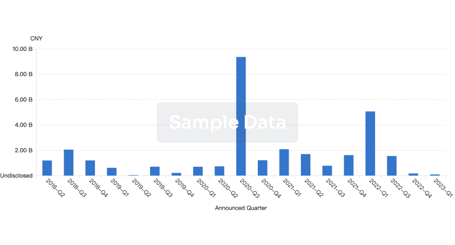
Financing
Unearth financing trends to validate and advance investment opportunities.
login
or
Chat with Hiro
Get started for free today!
Accelerate Strategic R&D decision making with Synapse, PatSnap’s AI-powered Connected Innovation Intelligence Platform Built for Life Sciences Professionals.
Start your data trial now!
Synapse data is also accessible to external entities via APIs or data packages. Empower better decisions with the latest in pharmaceutical intelligence.
Bio
Bio Sequences Search & Analysis
Sign up for free
Chemical
Chemical Structures Search & Analysis
Sign up for free